Nuclear medicine
Nuclear medicine is a specialized area of radiology that uses very small amounts of radioactive materials, or radiopharmaceuticals, to examine organ function and structure. Nuclear medicine imaging is a combination of many different disciplines.
Nuclear Medicine...
Nuclear medicine is a medical specialty that uses small amounts of radioactive material in the diagnosis and treatment of the disease in early-stage like heart disease, neurological disorders, several types of cancer and other abnormalities of the body. Nuclear medicine is a branch of medical imaging that uses small amounts of radioactive material to diagnose and determine the severity of or treat a variety of diseases, including many types of cancers, heart disease, gastrointestinal, endocrine, neurological disorders and other abnormalities within the body.
Nuclear medicine is used to diagnose a wide range of conditions.
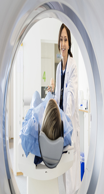
The patient will inhale, swallow, or be injected with a radiopharmaceutical. This is a radioactive material. After taking the substance, the patient will normally lie down on a table, while a camera takes pictures.
• How does Camera work?
The camera will focus on the area where the radioactive material is concentrated, and this will show the doctor what kind of a problem there is, and where it is.
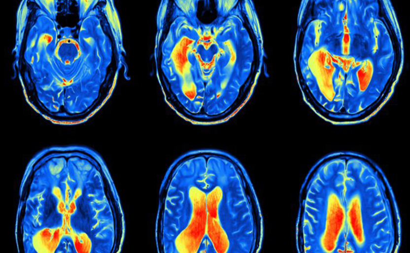
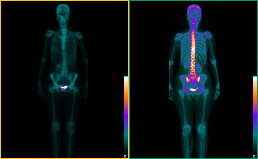
Types of imaging techniques include positon emission tomography (PET) and single-photon emission computed tomography (SPECT).
PET and SPECT scans can provide detailed information about how a body organ is functioning.
This type of imaging is particularly helpful for diagnosing thyroid disease, gall bladder disease, heart conditions, and cancer. It can also help diagnose Alzheimer's disease and other types of dementia and brain conditions.
In the past, diagnosing internal problems often needed surgery, but nuclear medicine makes this unnecessary.
After diagnosis, and when treatment starts, PET and SPECT can show how well the treatment is working.
PET and SPECT are also offering new insights into psychiatric conditions, neurological disorders, and addiction.
Other types of imaging involved in nuclear medicine include targeted molecular ultrasound, which is useful in detecting different kinds of cancer and highlighting blood flow; and magnetic resonance sonography, which has a role in diagnosing cancer and metabolic disorders.
• In the past...
Diagnosing internal problems often needed surgery, but nuclear medicine makes this unnecessary.
After diagnosis, and when treatment starts, PET and SPECT can show how well the treatment is working.
PET and SPECT are also offering new insights into psychiatric conditions, neurological disorders, and addiction.
Other types of imaging involved in nuclear medicine include targeted molecular ultrasound, which is useful in detecting different kinds of cancer and highlighting blood flow; and magnetic resonance sonography, which has a role in diagnosing cancer and metabolic disorders.
Radioactive agents, I will be swallowed in pill form, inhaled, or injected as part of a person's treatment.
Radioactive techniques are also used in treatment. The same agents that are used in nuclear imaging can be used to deliver treatment. The radiopharmaceutical can be swallowed, injected, or inhaled.
One example is radioactive iodine (I-131). It has been used for over 50 years to treat thyroid cancer and hyperthyroidism, or an overactive thyroid. Now, it is also used to treat non-Hodgkin lymphoma and bone pain from some kinds of cancer.
Iodine-131 (I-131) targeted radionuclide therapy (TRT) introduces radioactive iodine into the body. As the thyroid cells or cancer cells absorb this substance, it kills them. I-131 can be given as capsules or in liquid form.
Nuclear imaging...
The patient may have to wear a gown, or they may be able to wear their own clothes, but they will have to remove the jewelry and other metal-base accessories.
Therapy
After having radioactive treatment, a person should avoid physical contact with other people as much as possible for 2-5 days, which may involve taking time off work.
When a patient has treatment for the thyroid with I-131, no special equipment is used.
A single, prepared dose will be taken by mouth. This is a one-time treatment.
The patient should not eat or drink after midnight on the day of the treatment. If the treatment is for a thyroid problem, the doctor will normally advise them to stop taking their regular thyroid medication between 3 and 7 days before the treatment.
The patient may be able to return home after the dose, or they may have to stay overnight in the hospital.
However, because the body will not absorb all the radioactive iodine, it will continue to leave the body over the next 2 to 5 days.
The individual should avoid contact with other people as far as possible, and especially with infants and pregnant women.
This may mean taking time off work. They should also prepare their own food, avoid sleeping with another person, flush the lavatory twice after use, and wash their clothes and laundry separately.
Most of the iodine will leave the body through the urine, but it is also excreted through tears, sweat, saliva, vaginal discharge, and feces.
Women are advised to avoid becoming pregnant for 6 to 12 months following treatment.
Anyone who plans to travel immediately after treatment should get a letter from the doctor, as radioactivity may show up on scanning machines at airports.
Safety in nuclear medicine
Too much radiation can potentially damage organs or tissues or increase the risk of cancer.
However, when used for diagnosis, the level of radiation exposure is around the same as a person receives during a routine chest x-ray or a CT scan. As a result, nuclear medicine and imaging procedures are considered non-invasive and relatively safe. Their effectiveness in diagnosing disease means that the benefits normally outweigh the risks.
Treatment with nuclear medicine involves larger doses of radioactive material.
For example, a nuclear medicine lung scan would expose a person to 2 millisieverts (mSv) of radioactivity, while cancer treatment would expose a tumor to 50,000 mSv.
This additional dose may affect the patient, and side effects are possible.
However, since the treatment often targets potentially fatal diseases, the benefits tend to outweigh the risks.
As technology advances, scientists hope that treatments will be more directed toward the tumor or disease, and less likely to affect the person as a whole.
The Nuclear Regulatory Commission (NRC) and the U.S. Food and Drug Administration (FDA) closely regulate the use of radioactive materials for nuclear medicine to ensure the safety of patients.