Ultrasound
Ultrasound imaging uses sound waves to produce pictures of the inside of the body. ... What is General Ultrasound Imaging?... Ultrasound imaging is also called ultrasound scanning or sonography...
What is General Ultrasound Imaging?
Medical ultrasound is a diagnostic imaging technique based on the application of ultrasound. It is used to create an image of internal body structures such as tendons, muscles, joints, blood vessels, and internal organs. Its aim is often to find a source of a disease or to exclude pathology.
Ultrasound scans: How do they work?
• What is Medical Ultrasound?
• An ultrasound scan uses high-frequency sound waves to create images of the inside of the body. It is suitable for use during pregnancy.
• Ultrasound scans, or sonography, are safe because they use sound waves or echoes to make an image, instead of radiation.
• Ultrasound scans are used to evaluate fetal development, and they can detect problems in the liver, heart, kidney, or abdomen. They may also assist in performing certain types of biopsy. The image produced is called a sonogram.
Fast facts on ultrasound scans...
Here are some key points about ultrasound scans. More detail is in the main article.
• Ultrasound scans are safe and widely used.
- They are often used to check the progress of a pregnancy.
- They are used for diagnosis or treatment.
- No special preparation is normally necessary before an ultrasound scan.
• Ultrasound scans are carried out by a sonographer.
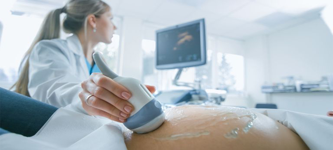
The person who performs an ultrasound scan is called a sonographer, but the images are interpreted by radiologists, cardiologists, or other specialists.
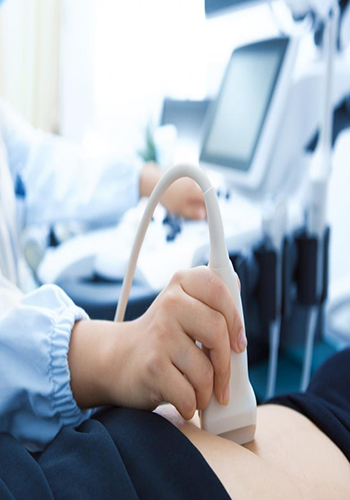
The sonographer usually holds a transducer, a hand-held device, like a wand, which is placed on the patient's skin.
Ultrasound is sound that travels through soft tissue and fluids, but it bounces back, or echoes, off denser surfaces. This is how it creates an image.
The sonographer usually holds a transducer, a hand-held device, like a wand, which is placed on the patient's skin.
Ultrasound is sound that travels through soft tissue and fluids, but it bounces back, or echoes, off denser surfaces. This is how it creates an image.
• Ultrasound
The term "ultrasound" refers to sound with a frequency that humans cannot hear.
For diagnostic uses, the ultrasound is usually between 2 and 18 megahertzTrusted Source (MHz).
Higher frequencies provide better quality images but are more readily absorbed by the skin and other tissue, so they cannot penetrate as deeply as lower frequencies.
Lower frequencies penetrate deeper, but the image quality is inferior.
• How does it capture an image?
Ultrasound will travel through the blood in the heart chamber, for example, but if it hits a heart valve, it will echo, or bounce back.
It will travel straight through the gallbladder if there are no gallstones, but if there are stones, it will bounce back from them.
The denser the object the ultrasound hits, the more of the ultrasound bounces back.
This bouncing back, or echo, gives the ultrasound image its features. Varying shades of gray reflect different densities.
Ultrasound transducers
The transducer, or wand, is normally placed on the surface of the patient's body, but some kids are placed internally.
These can provide clearer, more informative images.
• Examples are:
- an endovaginal transducer, for use in the vagina.
- an endorectal transducer, for use in the rectum.
- a transesophageal transducer passed down the patient's throat for use in the esophagus.
- Some very small transducers can be placed onto the end of a catheter and inserted into blood vessels to examine the walls of blood vessels.
- an endorectal transducer, for use in the rectum.
- a transesophageal transducer passed down the patient's throat for use in the esophagus.
- Some very small transducers can be placed onto the end of a catheter and inserted into blood vessels to examine the walls of blood vessels.
• Uses
Ultrasound images are made from reflected sound, and a diagnosis can then be made.
Ultrasound is commonly used trusted Source for diagnosis, for treatment, and for guidance during procedures such as biopsies.
It can be used to examine internal organs such as the liver and kidneys, the pancreas, the thyroid gland, the testes and the ovaries, and others.
Ultrasound is commonly used trusted Source for diagnosis, for treatment, and for guidance during procedures such as biopsies.
It can be used to examine internal organs such as the liver and kidneys, the pancreas, the thyroid gland, the testes and the ovaries, and others.
• Ultrasound scan
An ultrasound scan can reveal whether a lump is a tumor. This could be cancerous, or a fluid-filled cyst.
It can help diagnose problems with soft tissues, muscles, blood vessels, tendons, and joints. It is used to investigate a frozen shoulder, tennis elbow, carpal tunnel syndrome, and others.
It can help diagnose problems with soft tissues, muscles, blood vessels, tendons, and joints. It is used to investigate a frozen shoulder, tennis elbow, carpal tunnel syndrome, and others.
• An echocardiogram (ECG)
is an example of a Doppler ultrasound. It can be used to create images of the cardiovascular system and to measure blood flow and cardiac tissue movement at specific points.
A Doppler ultrasound can assess the function and state of cardiac valve areas, any abnormalities in the heart, valvular regurgitation, or blood leaking from valves, and it can show how well the heart pumps out blood.
A Doppler ultrasound can assess the function and state of cardiac valve areas, any abnormalities in the heart, valvular regurgitation, or blood leaking from valves, and it can show how well the heart pumps out blood.
• Circulatory problems
Doppler ultrasound can assess the flow of blood in a vessel or blood pressure. It can determine the speed of the blood flow and any obstructions.
• It can also be used to:
- examine the walls of blood vessels.
- check for DVT or an aneurysm.
- check the fetal heart and heartbeat.
- evaluate for plaque buildup and clots.
- assess for blockages or narrowing of arteries.
- A carotid duplex is a form of carotid ultrasonography that may include a Doppler ultrasound. This would reveal how blood cells move through the carotid arteries.
• Ultrasound in anesthesiology
Ultrasound is often used by anesthetists to guide a needle with anesthetic solutions near nerves.
An ultrasound can be done at a doctor's office, at an outpatient clinic, or in the hospital.
Most scans take between 20 and 60 minutes. It is not normally painful, and there is no noise.
In most cases, no special preparation is needed, but patients may wish to wear loose-fitting and comfortable clothing.
If the liver or gallbladder is affected, the patient may have to fast or eat nothing, for several hours before the procedure.
For a scan during pregnancy, and especially early pregnancy, the patient should drink plenty of water and try to avoid urinating for some time before the test.
When the bladder is full, the scan produces a better image of the uterus.
The scan usually takes place in the radiology department of a hospital. A doctor or a specially-trained sonographer will carry out the test.