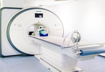
Mri scanner
Magnetic resonance imaging is a medical imaging technique used in radiology to form pictures of the anatomy and the physiological processes of the body. MRI scanners use strong magnetic fields, magnetic field gradients, and radio waves to generate images of the organs in the body.
Fast facts on MRI scanning:
• MRI scanning is a non-invasive and painless procedure.
• Raymond Damadian created the first MRI full-body scanner, which he nicknamed the Indomitable.
• The cost of a basic MRI scanner starts at $150,000 but can exceed several million dollars.
• Japan has the most MRI scanners per capita, with 48 machines for every 100,000 citizens.
• An MRI scan uses a large magnet, radio waves, and a computer to create a detailed, cross-sectional image of internal organs and structures.
• An MRI scan differs from CT scans and X-rays, as it does not use potentially harmful ionizing radiation.
This list is by no means exhaustive. The use of MRI technology is always expanding in scope and use.
Uses :
The development of the MRI scan represents a huge milestone for the medical world & Doctors, scientists, & researchers are now able to examine the inside of the human body in high detail using a non-invasive tool.
The following are examples in which an MRI scanner would be used:
Anomalies of the brain and spinal cord.
The evaluation of pelvic pain in women, with causes including fibroids and endometriosis.
Tumors, cysts, and other anomalies in various parts of the body.
Suspected uterine anomalies in women undergoing evaluation for infertility.
Injuries or abnormalities of the joints, such as the back and knee.
Diseases of the liver and other abdominal organs.
Breast cancer screening for women who face a high risk of breast cancer.
Certain types of heart problems.
A person can listen to music in headphones to mask the loud and sometimes alarming sound of the MRI machine.
There is very little preparation required, if any, before an MRI scan.
On arrival at the hospital, doctors may ask the patient to change into a gown. As magnets are used, it is critical that no metal objects are present in the scanner. The doctor will ask the patient to remove any metal jewelry or accessories that might interfere with the machine.
A person will probably be unable to have an MRI if they have any metal inside their bodies, such as bullets, shrapnel, or other metallic foreign bodies. This can also include medical devices, such as cochlear implants, aneurysm clips, and pacemakers.
Individuals who are anxious or nervous about enclosed spaces should tell their doctor. Often they can be given medication prior to the MRI to help make the procedure more comfortable.
Patients will sometimes receive an injection of intravenous (IV) contrast liquid to improve the visibility of a particular tissue that is relevant to the scan.
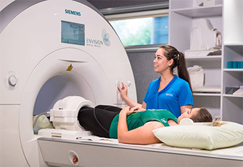
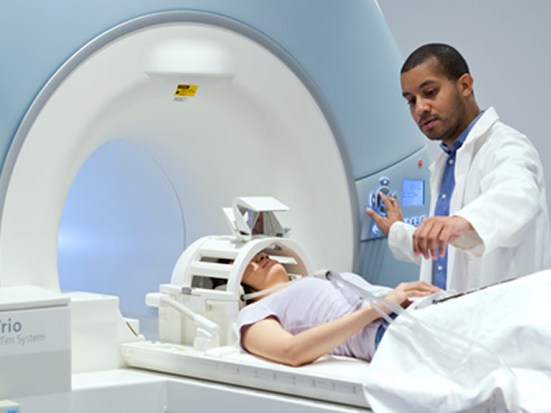
The radiologist, a doctor who specializes in medical images, will then talk the individual through the MRI scanning process and answer any questions they may have about the procedure.
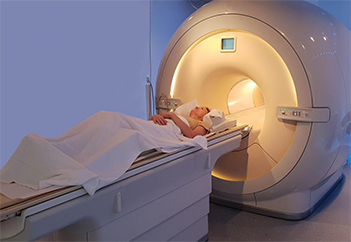
Once the patient has entered the scanning room, the doctor will help them onto the scanner table to lie down. Staff will ensure that they are as comfortable as possible by providing blankets or cushions.
Earplugs or headphones will be provided to block out the loud noises of the scanner. The latter is popular with children, as they can listen to music to calm any anxiety during the procedure.
There is very little preparation required, if any, before an MRI scan.
On arrival at the hospital, doctors may ask the patient to change into a gown. As magnets are used, it is critical that no metal objects are present in the scanner. The doctor will ask the patient to remove any metal jewelry or accessories that might interfere with the machine.
A person will probably be unable to have an MRI if they have any metal inside their bodies, such as bullets, shrapnel, or other metallic foreign bodies. This can also include medical devices, such as cochlear implants, aneurysm clips, and pacemakers.
Individuals who are anxious or nervous about enclosed spaces should tell their doctor. Often they can be given medication prior to the MRI to help make the procedure more comfortable.
Patients will sometimes receive an injection of intravenous (IV) contrast liquid to improve the visibility of a particular tissue that is relevant to the scan.
The radiologist, a doctor who specializes in medical images, will then talk the individual through the MRI scanning process and answer any questions they may have about the procedure.
Once the patient has entered the scanning room, the doctor will help them onto the scanner table to lie down. Staff will ensure that they are as comfortable as possible by providing blankets or cushions.
Earplugs or headphones will be provided to block out the loud noises of the scanner. The latter is popular with children, as they can listen to music to calm any anxiety during the procedure.
Once in the scanner, the MRI technician will communicate with the patient via the intercom to make sure that they are comfortable. They will not start the scan until the patient is ready.
During the scan, it is vital to stay still. Any movement will disrupt the images, much like a camera trying to take a picture of a moving object. Loud clanging noises will come from the scanner. This is perfectly normal. Depending on the images, at times it may be necessary for the person to hold their breath.
If the patient feels uncomfortable during the procedure, they can speak to the MRI technician via the intercom and request that the scan is stopped.
After the scan, the radiologist will examine the images to check whether any more are required. If the radiologist is satisfied, the patient can go home.
The radiologist will prepare a report for the requesting doctor. Patients are usually asked to make an appointment with their doctor to discuss the results.
It is extremely rare that a patient will experience side effects from an MRI scan.
However, the contrast dye can cause nausea, headaches, and pain or burn at the point of injection in some people. Allergy to the contrast material is also seldom seen but possible and can cause hives or itchy eyes. Notify the technician if any adverse reactions occur.
People who experience claustrophobia or feel uncomfortable in enclosed spaces sometimes express difficulties with undergoing an MRI scan.
MRI scans work by rearranging water molecules in the body with magnets.
An MRI scanner contains two powerful magnets. These are the most important parts of the equipment.
The human body is largely made of water molecules, which are comprised of hydrogen and oxygen atoms. At the center of each atom lies an even smaller particle called a proton, which serves as a magnet and is sensitive to any magnetic field.
Normally, the water molecules in the body are randomly arranged, but on entering an MRI scanner, the first magnet causes the water molecules to align in one direction, either north or south.
The second magnetic field is then turned on and off in a series of quick pulses, causing each hydrogen atom to change its alignment when switched on and then quickly switch back to its original relaxed state when switched off.
Passing electricity through gradient coils, which also causes the coils to vibrate, creates the magnetic field, causing a knocking sound inside the scanner.
Although the patient cannot feel these changes, the scanner can detect them and, in conjunction with a computer, can create a detailed cross-sectional image for the radiologist.
Functional magnetic resonance imaging (fMRI)
Functional magnetic resonance imaging or functional MRI (fMRI) uses MRI technology to measure cognitive activity by monitoring blood flow to certain areas of the brain.
• Helps
The blood flow increases in areas where neurons are active. This gives an insight into the activity of neurons in the brain.
This technique has revolutionized brain mapping, by allowing researchers to assess the brain and spinal cord without the need for invasive procedures or drug injections.
Functional MRI helps researchers learn about the function of a normal, diseased, or injured brain.
• Used
fMRI is also used in clinical practice. Standard MRI scans are useful for detecting anomalies in tissue structure. However, an fMRI scan can help detect anomalies in the activity.
In short, fMRI tests what tissues do rather than how they look.
As such, doctors use fMRI to assess the risks of brain surgery by identifying the regions of the brain involved in critical functions, such as speaking, movement, sensing, or planning.
Functional MRI can also be used to determine the effects of tumors, stroke, head and brain injuries, or neurodegenerative diseases, such as Alzheimer's.